Download A4Medicine Mobile App
Empower Your RCGP AKT Journey: Master the MCQs with Us!

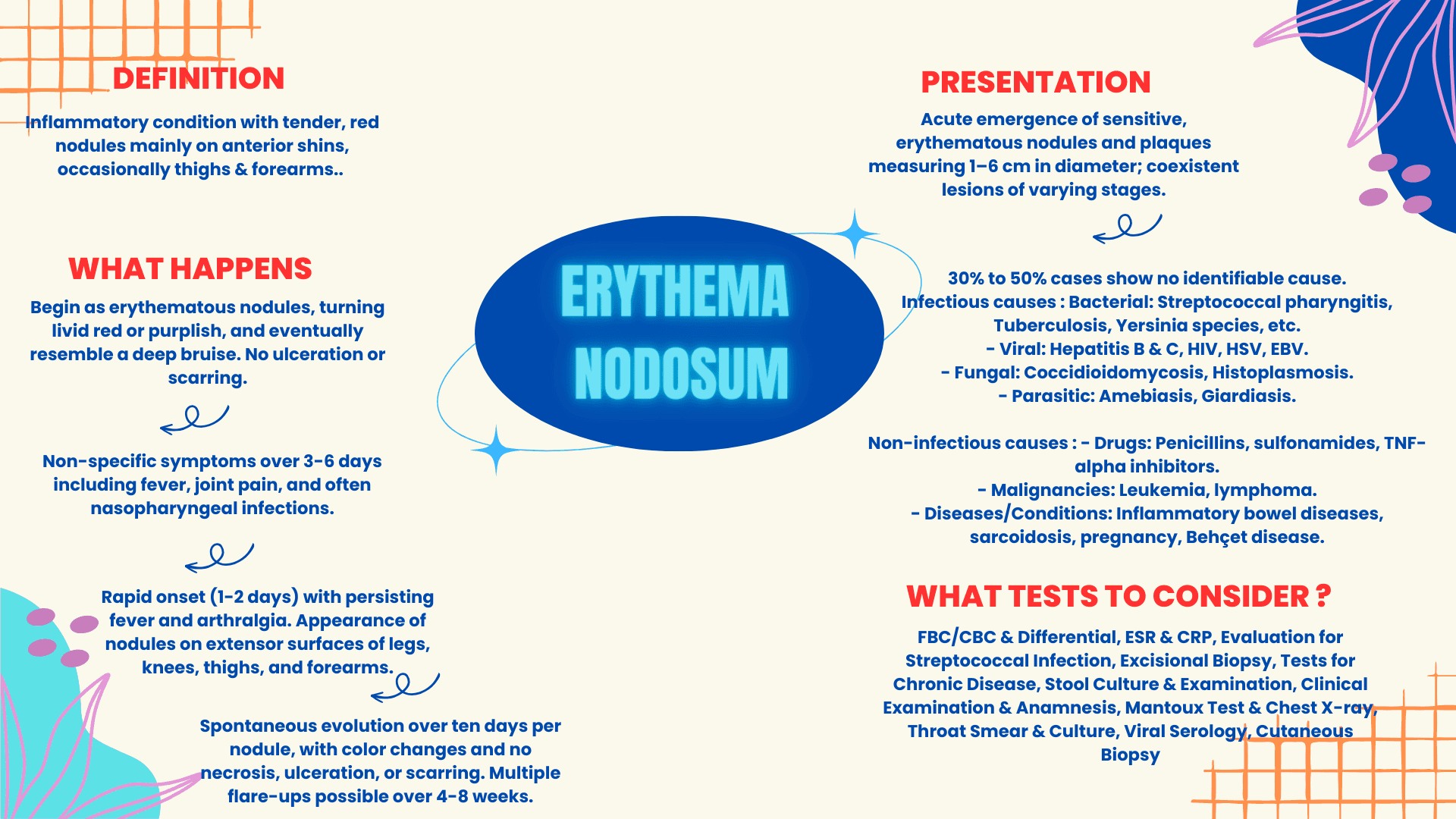
This table offers a comprehensive overview of Erythema Nodosum, listing its clinical presentations, evolutionary stages, potential causes, and primary diagnostic measures. By amalgamating key features and associated conditions, it serves as a succinct reference for clinicians and medical students to understand this inflammatory skin condition and its broader implications.
| Feature | Description/Examples |
|---|---|
| Basic Description | Inflammatory condition with tender, red nodules mainly on anterior shins, occasionally thighs & forearms. |
| Histopathology | Mainly septal panniculitis without vasculitis. |
| Evolution of Nodules | Begin as erythematous nodules, turning livid red or purplish, and eventually resemble a deep bruise. No ulceration or scarring. |
| Prodromal Phase | Non-specific symptoms over 3-6 days including fever, joint pain, and often nasopharyngeal infections. |
| Stade Phase | Rapid onset (1-2 days) with persisting fever and arthralgia. Appearance of nodules on extensor surfaces of legs, knees, thighs, and forearms. |
| Regressive Phase | Spontaneous evolution over ten days per nodule, with color changes and no necrosis, ulceration, or scarring. Multiple flare-ups possible over 4-8 weeks. |
| Clinical Presentation | Acute emergence of sensitive, erythematous nodules and plaques measuring 1–6 cm in diameter; coexistent lesions of varying stages. |
| Idiopathic Cases | 30% to 50% cases show no identifiable cause. |
| Infectious Causes | - Bacterial: Streptococcal pharyngitis, Tuberculosis, Yersinia species, etc. - Viral... |
Try our Free Plan to get the full article.